
Recovery and resilience plan.
The most common method to quantify the plasma effect on biofilm is to scrape biofilm from the support medium to a solution and to vortex or directly sonicate biofilm with its support in order to release individual bacteria into a solution. Then serial dilutions suspension with bacteria take place and subsequently plating on solid/agar media and cultivation and finally counting grown colonies. However in a biofilm, bacteria are in various metabolic states. During plasma exposure they are introduced into a new environment with changing chemical conditions, temperature, pH, electric fields which can induce enzyme synthesis and adjustments of metabolic pathways. Therefore bacterial cells after plasma treatment could be intact and alive, according to metabolic criteria, but do not undergo cell division on a bacteriological medium, we call these bacteria viable but non-culturable. Another reason why cultivation of cells from biofilm can skew results is that bacteria in biofilms are embedded in polymeric substance. It protects them from the environment, thus disruption of this substance can be really difficult, not all bacteria are released to the liquid medium, and during sonication some of cells could be disrupted by shock waves. As a result, the method of cultivation shows only cultivable/culturable bacteria which undergo a cell division sufficient number of times to form a visible colony on agar surface.
Another method very often used especially for qualitative observation of biofilm is LIVE/DEAD Viability Kit (Invitrogen). It provides a two-color fluorescence assay of bacterial viability, based on membrane integrity and utilizes mixtures of the SYTO 9 green fluorescent nucleic acid stain and the red fluorescent nucleic acid stain, propidium iodide (PI). These stains differ both in their spectral characteristics and in their ability to penetrate healthy bacterial cells. When used alone, the SYTO 9 stain generally labels all bacteria in a population - those with intact membranes and those with damaged membranes. In contrast, PI penetrates only bacteria with damaged membranes, causing a reduction in the SYTO 9 stain fluorescence when both dyes are present. Thus, with an appropriate mixture of the SYTO 9 and PI stains, bacteria with intact cell membranes (live) stain fluorescent green, whereas bacteria with damaged membranes (dead) stain fluorescent red. The excitation/emission maxima for these dyes are approximately 480/500 nm for SYTO 9 and 490/635 nm for PI. The background remains virtually non-fluorescent. This method was used in studies mostly for qualitative observation, although can also be used for quantification. However, the plasma treatment can destroy cell parts or even entire cells and if there is not enough membrane preserved to concentrate PI to label the cells as dead, then the live/dead fluorescence ratio can be shifted towards live. On the other hand, PI can in some cases penetrate into living cells with small membrane punctures which could have been easily repaired, but LIVE/DEAD stain will mark these cells as dead.
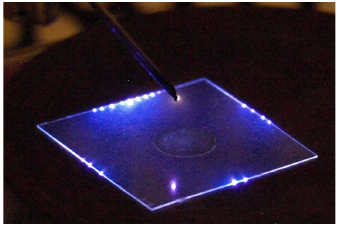
In our experiments we used both methods to evaluate impact on 48 hour old Escherichia coli biofilm on cover slides (2×2 cm) - cultivation and confocal laser scanning microscopy. Fluorescent stains BacLight LIVE/DEAD and DAPI were used for to evaluate ration of live and dead bacteria and to localize all DNA in samples and thus see changes in thickness of biofilm, respectively. The slides were analysed with inverted confocal laser scanning microscope Leica SPX8 (Imagif - CNRS, Gif-sur-Yvette, France) and Olympus IX81 (Department of Genetics, Faculty of Natural Sciences, Comenius University Bratislava, Slovakia). Both discharge setups - corona discharge in air (positive streamer corona and negative Trichel pulses) and DBD argon jet in argon atmosphere significantly reduced viability and thickness (reduction by 2/3) of biofilm within 20 min. By thermostatic cultivation of scraped biofilm solution we were able to reach 6 order of magnitude reduction of bacterial population in biofilm by applying argon jet for 20 min and 5 orders of magnitude reduction for both coronas in air (Kovalova Z. et al., 2014).

Recovery and resilience plan.
Old website of our division.
In 2022 we organised 9th Central European symposium on Plasma Chemistry and COST Action CA19110 workshop.
In 2016 we organised 6th International Conference on Plasma Medicine, with almost 400 participants.
Do not hesitate to contact us, if needed.