
Recovery and resilience plan.

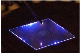
In collaboration with our Romanian partner (University of Iasi) we tested viability, apoptosis and cell cycle of normal (Vero) and cancerous (HeLa) mammalian cells (Hensel et al., 2015). Figure 1 shows the results of the direct TS treatment of HeLa cells. The initial concentration of the cells was approximately 5×10⁵ cells/mL in 5 mL of PBS (buffered solution of NaCl in deionized water).
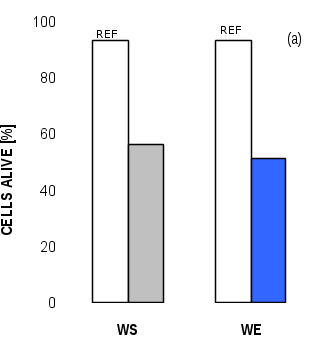
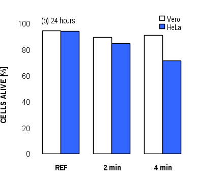
Figure 2 shows the viability of Vero and HeLa cells indirectly exposed to the plasma for 2 and 4 minutes after 4 and 24 hours of subsequent incubation. The incubation is the time interval the cells were allowed to grow in an incubator after the plasma exposure to enter to the log phase. After that, the cells were detached, re-suspended in PBS and their number determined by propidium iodide flow cytometry. The total cytotoxic effect of indirect exposure was much smaller compared to the direct exposure. The cytotoxic effect increased with the treatment time. While for HeLa cells the cytotoxic effect observed after 4 and 24 hours of incubation remained relatively stable (Fig. 2), in the case of Vero cells the cytotoxic effect was significantly attenuated with prolongation of the incubation period to 24 hours. The result indicates that the cancerous cells can be selectively targeted and killed as they are more sensitive to the plasma exposure, while normal cells are more resistant to external injuries and more easily able to repair eventual damages.
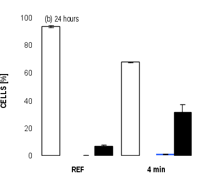
In order to probe the cell behavior before their death, the test of cell apoptosis was performed. The apoptosis was analyzed by apoptotic assay utilizing bivariate staining analysis by Annexin V-FITC and propidium iodide. Annexin V-FITC was used to identify the preapoptotic and apoptotic behavior, while propidium iodide was used to distinguish between live and dead cells. Figure 3 shows the results of the test performed on HeLa cells with indirect TS plasma exposure and for different incubation times. The analysis of the cells exposed to plasma and incubated for 4 hours showed the number of cells that became apoptotic (27%). The analysis of the cells incubated for 24 hours showed apoptotic cells disappearing from cell culture due to high disintegration of their structures and the corresponding increase of dead cells (32%) as a result of defective function of reparatory mechanisms in cancerous cells. Non affected cells or the remaining cells are able to proliferate, especially in the case of cancerous cells where proliferation is very intense. Also, depending on the moment of the cell cycle, some cells can trigger apoptosis, while others are not affected (especially in quiescent phase of cell cycle). Compared with HeLa cells, normal Vero cells are less sensitive to plasma exposure and the occurrence of small number of dead cells after 24 hours suggests that the part of Vero cells that were damaged by plasma were eliminated while a fraction of cells followed reparatory mechanisms.

Higher susceptibility of cancerous cell to plasma treatment was confirmed by the cell cycle analysis. Due to high proliferation rate, HeLa cells are more sensitive to plasma because a higher number of cells are in S phase. Overall damage impact is higher and prolonged in time. Not all damages occur immediately after plasma exposure but could be generated by secondary metabolites or other mechanisms.
Besides mammalian cells, we tested the potential plasma-induced apoptotic pathways on yeast cells (Saccharomyces cerevisiae) in aqueous suspension directly or indirectly exposed to the air plasma of transient spark with electrospray. Interestingly, yeast mutants defective in enzymes employed in yeast apoptosis demonstrated very small changes of survival rate in comparison with standard strains. The apoptotic pathways induced by plasma in model yeast cells were thus not clearly confirmed.
On the other hand, yeast mutants defective in synthesizing the superoxide dismutase enzyme as a defense against oxidative stress (BY4741Δsod1 and BY4741Δsod2) were found much more susceptible to plasma treatment, both direct and indirect. Longer incubation in the plasma treated solution results in lower survival rate of the cells, indicating the dominant role of the long-lived RONS in water created during the plasma treatment, which is in agreement with the bactericidal effects of plasma treated water.
Recovery and resilience plan.
Old website of our division.
In 2022 we organised 9th Central European symposium on Plasma Chemistry and COST Action CA19110 workshop.
In 2016 we organised 6th International Conference on Plasma Medicine, with almost 400 participants.
Do not hesitate to contact us, if needed.